
The Science · July 22, 2026 · 7 min · By Kofi Adusei
Fat Necrosis After a BBL: What Those Hard Lumps Are and When to Worry
Weeks after surgery, a firm nodule appears under the skin. Most of the time it is grafted fat that did not survive, and it is harmless. Knowing which lumps are normal and which are not is the difference between waiting it out and calling your surgeon tonight.
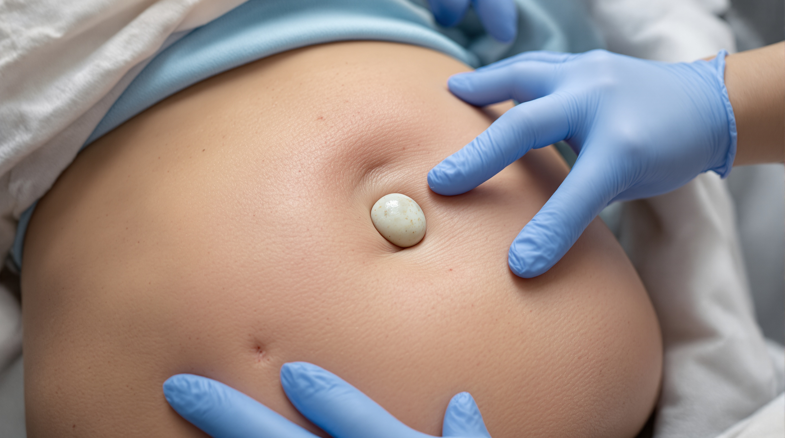
Somewhere between week three and week eight after a BBL, a lot of patients find a lump. It is usually firm, sometimes tender, often about the size of a marble or a small grape, and it was not there the week before. The internet immediately supplies the worst possible explanations. The far more likely answer is fat necrosis: a pocket of transferred fat that did not establish a blood supply, died, and is now being slowly cleared and walled off by the body. It is the most common structural complication of any fat grafting procedure, it is usually benign, and it has a predictable natural history. Here is what is actually happening under the skin, and where the line sits between a normal lump and a call to your surgeon.
Why grafted fat dies in the first place
A BBL moves living fat cells from one part of your body to another. Those cells arrive with no blood supply of their own. For the first few days they survive on diffusion, absorbing oxygen and nutrients from the surrounding tissue fluid, and they only stabilize once new capillaries grow in and reconnect them. That process, called revascularization, is the entire game, and it is why surgeons inject fat in thin, widely dispersed threads rather than in a single large deposit. A thread of fat is close enough to living tissue at every point to be rescued. A bolus is not: the outside of the deposit revascularizes, the center is too far from a blood supply and dies.
A review in the journal Medicine describes fat necrosis as one of the most common complications after fat grafting and links it directly to inadequate blood supply and the volume and distribution of the graft (PMC, NIH). This is the same physiology that drives the wide variation in how much transferred fat actually survives, and it is why surgeons who overfill aggressively to chase a bigger result are also the ones whose patients report more nodules.
What the lump actually is
When a pocket of fat cells dies, the cell membranes rupture and release their oil. The body treats that free oil as a foreign substance and mounts an inflammatory response around it. Over weeks, immune cells wall the area off with a fibrous capsule. Depending on how much oil is involved, the result is either a firm scar-like nodule of fibrotic tissue or a soft, fluid-filled oil cyst. Some of these calcify over months or years, which is why they can show up as small dense spots on later imaging. A case report on granulomatous reaction after gluteal fat transfer documents exactly this sequence of walled-off, inflamed graft material presenting as palpable buttock nodules (PMC, NIH).
So the lump you are feeling is generally not new tissue and not a tumor. It is the remains of graft that did not take, plus the capsule your body built around it.
The normal timeline
Fat necrosis nodules rarely announce themselves in the first two weeks, because swelling masks everything. They typically become palpable between weeks three and eight, as the swelling recedes and the underlying architecture becomes readable through the skin. Small nodules, under a centimeter or so, frequently soften and shrink on their own over three to six months as the body reabsorbs the oil and remodels the fibrous tissue. Larger ones may persist and stay palpable permanently, though they usually become less noticeable as surrounding tissue settles. Our week-by-week recovery guide maps this against the rest of the healing timeline, and it is worth reading before you conclude that something has gone wrong.
Warning signs that are not fat necrosis
This is the part worth memorizing. A benign necrosis nodule is firm, well localized, and unaccompanied by systemic symptoms. Call your surgeon promptly, or seek emergency care, if the lump comes with any of the following: expanding redness or heat over the skin, fever or chills, drainage of pus or foul-smelling fluid, rapidly increasing size, severe or escalating pain, or skin that darkens and breaks down over the area. Those describe infection or an abscess, not resorbed graft, and they need antibiotics or drainage rather than patience. Sudden shortness of breath or chest pain in the first days after surgery is a separate emergency entirely and is unrelated to nodules. The American Society of Plastic Surgeons is blunt that buttock fat grafting carries meaningfully more risk than most elective aesthetic procedures, which is exactly why symptoms outside the expected pattern deserve a same-day phone call rather than a forum search (ASPS).
How it gets treated
Most nodules need nothing. Time, gentle pressure, and patience resolve the small ones. Lymphatic massage performed by someone trained in post-surgical work can help soften fibrotic areas once your surgeon clears you for it, though it will not dissolve an established oil cyst. For persistent nodules, options range from ultrasound-guided aspiration of a fluid-filled cyst to a small in-office steroid injection to soften fibrosis, up to surgical excision of a stubborn firm nodule, usually deferred until at least six months out so the tissue has fully declared itself. Any nodule that is genuinely uncertain gets imaged first.
The prevention side is mostly the surgeon's job
You cannot control most of the variables that determine whether fat necrosis happens. Graft handling, injection technique, the size of the threads laid down, and above all the total volume forced into a given space are decided in the operating room. Patients who insist on maximum projection are asking for exactly the overfilling that produces nodules, which is one more reason the honest conversation about realistic volume matters. What you can control is who holds the cannula, which is the entire argument for vetting a surgeon's credentials and technique properly. After surgery, following the compression and positioning instructions protects perfusion in the graft while it is most vulnerable.
The takeaway
A firm lump appearing a month after a BBL is common, usually harmless, and usually the physical evidence of fat that did not survive the transfer. Judge it by the company it keeps: an isolated firm nodule with no fever, no spreading redness, and no drainage is almost always fat necrosis that time will improve, while a lump accompanied by heat, fever, pus, or rapid growth is an infection and needs your surgeon today, not next week. When in doubt, send a photo and get it looked at. Nodules are cheap to evaluate and expensive to ignore.
Related reading: Why fat survival rate after BBL varies so widely from patient to patient and BBL recovery, week by week.